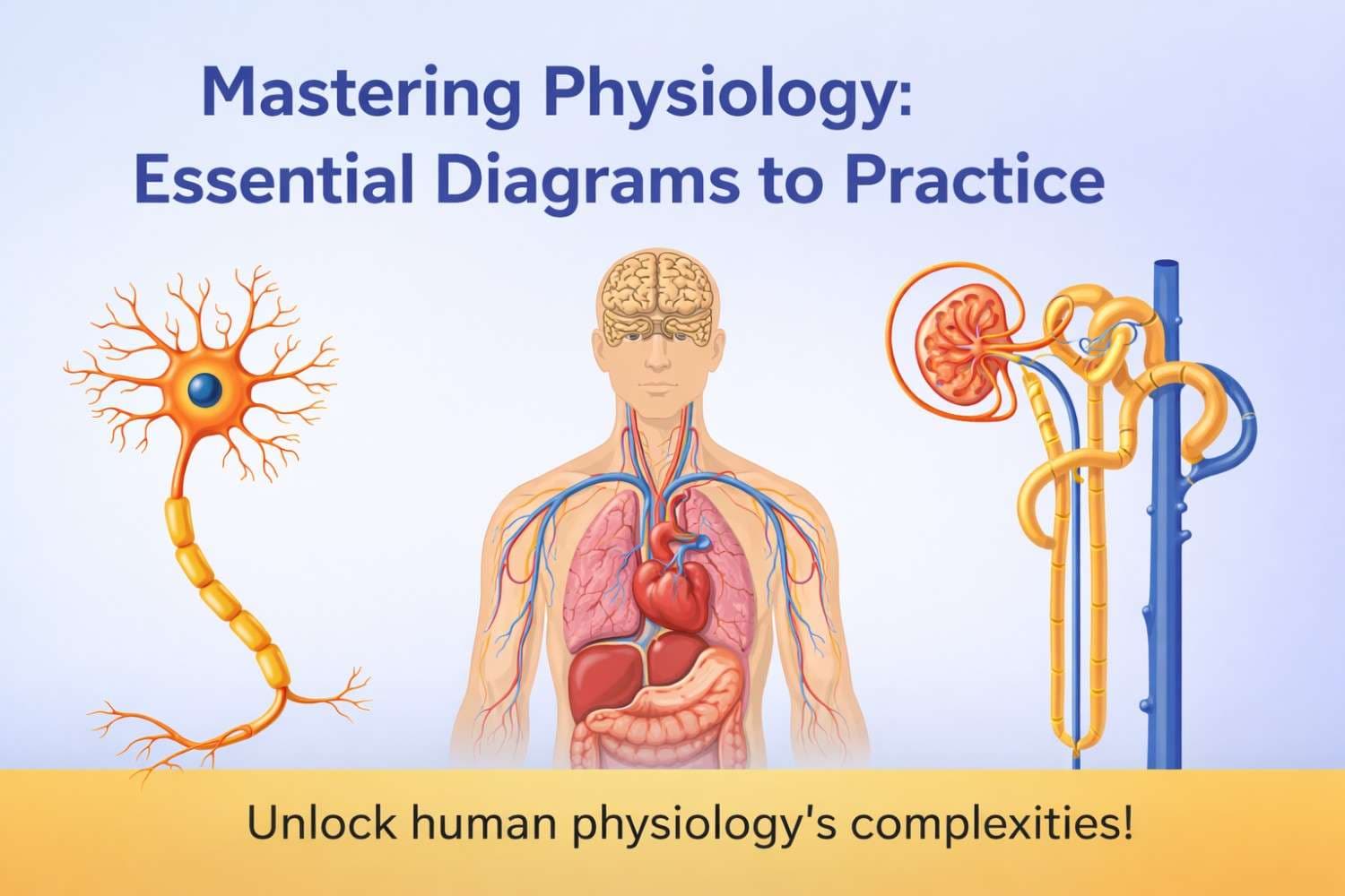
Mastering Physiology: Essential Diagrams to Practice
Table of Contents
Human physiology is a fascinating yet intricate subject, delving into the sophisticated mechanisms that keep our bodies functioning. While textbooks and lectures provide a wealth of information, one of the most powerful tools for truly grasping these complex processes is the humble diagram. Visual representations transform abstract concepts into tangible, understandable pathways and structures. Practicing these diagrams isn't just about memorization; it's about understanding connections, tracing flows, and solidifying your knowledge.
Why Diagrams are Indispensable
Diagrams serve as visual blueprints, allowing you to:
- Visualize Structures: Clearly see the anatomical components involved in a physiological process.
- Trace Pathways: Follow the flow of blood, nerve impulses, hormones, or substances through the body.
- Understand Relationships: Recognize how different organs, cells, and molecules interact.
- Consolidate Information: Bring together disparate pieces of knowledge into a coherent whole.
- Prepare for Exams: Many physiology questions involve labeling diagrams or explaining processes using visual aids.
Ready to boost your physiological understanding? Here are some of the most important diagrams you should actively practice:
The Nervous System: The Body's Electrical Network
1. The Neuron Structure
Understanding the components of a neuron (dendrites, cell body, axon, myelin sheath, synaptic terminals) is fundamental to comprehending how nerve impulses are generated and transmitted. Practice labeling and understand the function of each part in relation to action potentials and signal propagation.
2. The Synapse
The junction where one neuron communicates with another (or an effector cell) is critical. Diagramming the presynaptic terminal, synaptic cleft, and postsynaptic membrane, along with the release and reception of neurotransmitters, demystifies neural communication and drug actions.
3. The Reflex Arc
A simple yet powerful representation of neural pathways, a reflex arc (receptor, sensory neuron, integration center, motor neuron, effector) illustrates the basic mechanism of involuntary responses and provides a template for understanding more complex neural circuits.
The Cardiovascular System: Life's Pumping Engine
1. The Heart Structure & Blood Flow
Labeling the four chambers, valves, and major blood vessels (aorta, vena cavae, pulmonary artery/veins) is essential. More importantly, practice tracing the path of blood through the heart and lungs, understanding oxygenation and deoxygenation cycles.
2. The ECG Waveform
Learn to identify the P wave, QRS complex, and T wave, and connect each to specific electrical events in the cardiac cycle (atrial depolarization, ventricular depolarization, ventricular repolarization). This is key for understanding heart rhythm and function.
The Respiratory System: The Breath of Life
1. Alveolus and Capillary Exchange
This diagram showcases the crucial interface where gas exchange occurs. Practice drawing the thin barrier between the alveolar air and the pulmonary capillary, illustrating the diffusion of oxygen and carbon dioxide. Understand factors affecting this exchange.
The Urinary System: The Body's Filter
1. The Nephron Structure
The functional unit of the kidney, the nephron (glomerulus, Bowman's capsule, proximal convoluted tubule, loop of Henle, distal convoluted tubule, collecting duct), is complex but vital. Labeling each segment and understanding its role in filtration, reabsorption, and secretion is paramount for grasping urine formation and fluid balance.
2. Countercurrent Mechanism
While challenging, visualizing the countercurrent multiplier (in the loop of Henle) and exchanger (in the vasa recta) is crucial for understanding how the kidney concentrates urine. Focus on the movement of water and solutes.
The Endocrine System: Hormonal Orchestration
1. Hypothalamic-Pituitary Axis
This central regulatory pathway controls many endocrine glands. Diagramming the connection between the hypothalamus, anterior pituitary (via portal system), and posterior pituitary (via neural connections), along with the hormones involved, helps clarify endocrine control.
2. Specific Feedback Loops
Practice diagrams illustrating negative feedback loops, such as the regulation of thyroid hormones or blood glucose levels (involving insulin and glucagon). These diagrams highlight homeostatic control mechanisms.
The Muscular System: Movement & Power
1. The Sarcomere and Sliding Filament Model
Delve into the microscopic structure of a muscle fiber. Diagramming a sarcomere (actin, myosin, Z-discs, H-zone, A-band, I-band) and illustrating the sliding filament mechanism during contraction is essential for understanding muscle physiology.
General Tips for Diagram Practice
- Draw from Scratch: Don't just label; try to recreate diagrams from memory.
- Label Everything: Be meticulous with your labels, including arrows to indicate flow or direction.
- Add Explanations: Next to each part or arrow, briefly write down its function or what's happening.
- Color-Code: Use different colors for different substances, structures, or pathways.
- Connect to Function: Always ask yourself "What is this part doing?" or "How does this pathway contribute to overall body function?"
- Use Blank Diagrams: Many textbooks and online resources offer blank diagrams for practice.
Conclusion
Diagrams are not merely supplementary; they are foundational to mastering human physiology. By actively engaging with these visual aids, drawing them, labeling them, and understanding the processes they represent, you'll build a robust and lasting understanding of how the human body works. So, grab your pens and start drawing – your physiological knowledge will thank you for it!